

Misty J. Williams-Fritze, Benny Muraj, Amanda McSweeney, Fernando Garcia-Polite, Raffaele Melidone, Jonathon Kirk, Rami Tzafriri , Cyrille Sage. Cochlear Implant: Mini Swine or Sheep? Such a Tricky Choice.
Background: Cochlear implants (CI) are a growing market currently valued at $1.67 billion and projected to grow 10.6%/year from 2020 to 2027 (Figure 1). 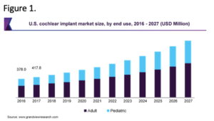 Since the first implantation in 1961, CI have undergone continuous technical improvements in sound processing, requiring associated safety testing in animals. While small animals provide a cost effective preclinical model for assessing hearing, their anatomical dimensions are not representative of the human, creating a need for large animal models.
Methods: We therefore investigated miniature swine and sheep as models for cochlear implantation using histology, endoscopy and functional readouts. Histology revealed round window membrane thickness to be species dependent, with the ovine anatomy more closely mimicking the human as compared to mini swine. As these differences are irrelevant for CI studies, we developed species specific endoscopic guided surgical procedures for accessing swine and ovine round windows. Cochlear implantation was performed on 3 mini swine (5-10 kg) through a posterior mandibular approach using the paracondylar process as the anatomical point of reference. The middle ear/round window was accessed by drilling at the edge of the auditory canal at a 45˚ angle. Implantation was verified perioperatively using CT scan (Figure 2).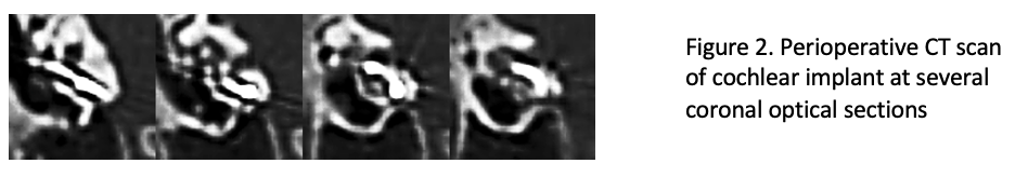
Results: ABR recordings were performed at 5 frequencies (2, 4, 8, 12 and 16 kHz) pre-implantation (baseline), and 30, 60 and 90 days post-implantation using an in-house custom-made ABR system that, unlike standard ABR systems, is sufficiently sensitive to acquire readouts from a standalone subcutaneous electrode. Evaluation of the un-implanted ear showed no sign of hearing deficiency at D90 compare to baseline, whereas the implanted ear showed a threshold shift of 40 to 50 dB at all frequencies. Following necropsy, the implanted ear was processed for histopathology using resin embedding, micro-grinding and eosin y-toluidine followed by pathologist assessment. No clearly visible inflammatory changes or fibrosis were identified in the examined sections in any of the implanted cochlea. The un-implanted cochlea were processed for cytocochleogram. Max projection images obtained from confocal scanning were fed to a customized image processing program for an efficient hair cell count of the unusually large samples. No hair cell loss was reported at the 5 frequencies tested during ABR (Figure 3), confirming the absence of processing artifacts.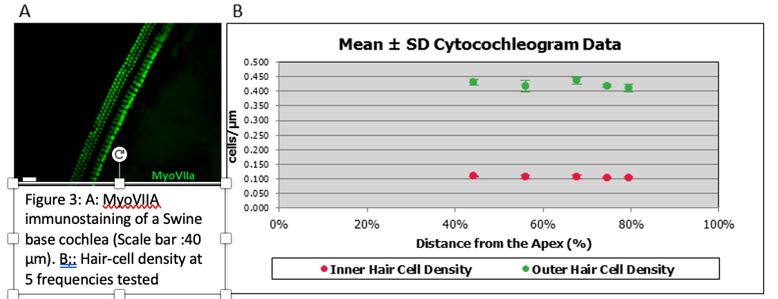
Conclusions: Taken together, these studies provide the methodology for a cochlear implantation in mini swine, from image-guided surgery, through functional readouts and down to immunohistochemical endpoint readouts such as inflammation and cell death. This methodology can be adapted to sheep, with some preliminary results already in place.
Presented at the Association for Research in Otolaryngology (ARO) 44th Annual Midwinter Virtual Meeting, Feb 20-24, 2021.