Supporting the Research Continuum, from Pilot to Regulatory Submission
Histology/histopathology is a critical endpoint for the non-clinical evaluation of medical devices, drugs, biologics and biomaterials. As a multidisciplinary research institute, CBSET supports a range of applications in therapeutic and device development, with AAALAC-accredited animal research facilities and GLP-compliant analyses.
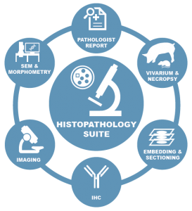
CBSET’s Comprehensive Histopathology Suite
Our histopathology suite and ACVP boarded pathologists provide flexible support and active collaboration for all facets of sample processing, staining, imaging and analysis, pairing it with expert interpretation and reporting. We will devise the right histopathology protocol for your aims and apply the optimal staining paradigm to satisfy your scientific endpoints.
Slide Preparation
Slide preparation capabilities include:
- Tissue trimming and fixation
- Paraffin and plastic resin processing
- Embedding, sectioning and staining
- Microtomy and EXAKT micro-grinding
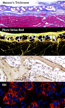
Staining Paradigms Selected to Elucidate Disease, Injury or Toxic Effect in Tissue
Primary staining provides rapid, low-cost visualization of any tissue type for robust qualitative assessments of histological changes. Common primary stain paradigms include:
- Cytology – e.g., Wright-Giemsa
- Histology
- Fibrosis/ repair – e.g., Masson’s trichrome
- Bone/ collagen – e.g., von Kossa, Goldner’s trichrome, safranin O
- Vascular – e.g., Movat’s pentachrome, van Gieson elastin
Secondary staining (IHC, IFA, ISH) is used for pinpoint contrast and quantitative analysis. Immunohistochemistry (IHC) and Immunofluorescence (IFA) use antibodies targeting cellular biomarkers, coupled to chromogenic enzymes or fluorophores, for improved dynamic range and sensitivity. In Situ Hybridization (ISH) detects expression of RNA in sections via oligonucleotide hybridization to target RNA sequences.
- Neurologic – e.g., neurofilament (NF), tyrosine hydroxylase (TH), calcitonin gene related peptide (CGRP)
- Connective tissue/vascular – e.g., collagen I, collagen III, von Willebrand factor, actin
- Inflammation – e.g., CD3, CD79a, C18, CD68
- Cell death and proliferation – e.g., TUNEL, proliferating cell nuclear antigen (PCNA)
- Gene expression (custom oligonucleotide probes directed to mRNA targets)
Tissue Biomarker Analysis
Tissue biomarker analysis can include:
- Method development
- Secondary probe strategies for optimized signal detection and amplification
- A wide range of fluorophores, or fluorophore-conjugated secondary antibodies
- Multiplexing for simultaneous visualization of multiple markers
- Quantitative analysis/ scoring of localization and intensity of signals
Expert Pathologist Assessment
CBSET can support your next study with industry-experienced, board-certified veterinary pathologists co-located with an integrated scientific team, to inform your regulatory and scientific endpoints with confidence.
- Written reports (GLP and non-GLP)
- Scoring paradigms and quantitative analysis
- Morphometry and SEM
 Watch: CBSET's John Keating presents In Vivo Models to Assess Ototoxicity