Cohen-Mazor M, Mathur P, Stanley JR, Mendelsohn FO, Lee H, Baird R, Zani BG, Markham PM, Rocha-Singh K. Evaluation of renal nerve morphological changes and norepinephrine levels following treatment with novel bipolar radiofrequency delivery systems in a porcine model.
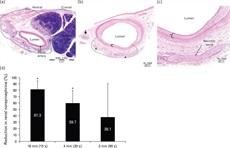
Summary: A porcine model (N = 46) was used to investigate renal norepinephrine levels and changes to renal artery tissues and nerves following percutaneous renal denervation with radiofrequency bipolar electrodes mounted on a balloon catheter. Percutaneous renal artery application of bipolar radiofrequency energy demonstrated safety and resulted in a significant renal norepinephrine content reduction and renal nerve injury compared with untreated controls in porcine models.

Electrode length studies. (a) Representative image of renal artery and surrounding tissues 7 days following 16-mm constant impedance radiofrequency treatment (75°C for 15 s). Note circumferential radiofrequency treatment of artery, as evidenced by transmural coagulative necrosis of the arterial wall and extension of thermal injury into the adjacent adventitia (i.e. zone of treatment within the black line) with affected nerves (arrowheads). (b) Representative image of renal artery and surrounding tissues 7 days following 4-mm constant impedance radiofrequency treatment (65°C for 30 s). Note localized radiofrequency treatment of artery, as evidenced by transmural coagulative necrosis of the arterial wall [in bracket and (c)] and extension of thermal injury into the adjacent adventitia (i.e. zone of treatment within dashed line) with affected nerves (arrowheads). (d) Mean (SD) percentage reduction of renal norepinephrine levels in the treated vs. untreated contralateral kidney 7 days following treatment with bipolar electrodes of length 16, 4, or 2 mm.*Statistically significant difference at P < 0.05 between RF-treated artery vs. untreated contralateral control. RF, radiofrequency.
J Hypertens. 2014 May 28. Full article download available here.

